nav
whatsapp icon

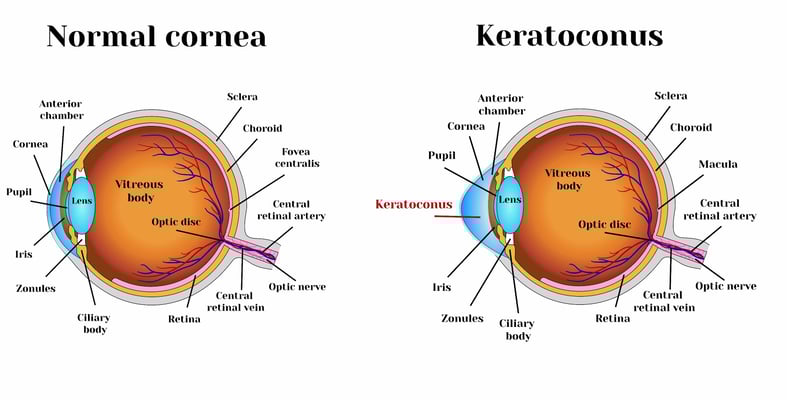
Keratoconus and cataracts , symptoms and types

blog single image

What is Keratoconus, How it looks and what are the symptoms? Also, find out Keratoconus’s types and stages , Learn more with ILAJAK Medical.

What is Keratoconus?

Keratoconus is when the cornea becomes thin and gradually bulges outward into a cone shape. Changing the shape of the cornea makes light rays out of focus, and as a result, your vision is blurry and distorted. This may make daily tasks such as reading or driving more difficult. Many keratoconus patients don't realize they have the disease.

Ilajak Medical offers the latest diagnostic techniques by the most skilled experts in Opthalmology.

Keratoconus diagnosis criteria

In addition to a full medical history and eye examination, an eye care specialist may perform the following tests for the diagnosis of keratoconus:

  • Corneal topography: This is the most accurate way to diagnose early keratoconus and follow its progression. A computerized image creates a map of the curve of the cornea.

Essentially, corneal topography measures the elevation of the cornea similar to geographic topography that shows hills and valleys on a map.  Topography is color-coded with cooler colors (blues) being flatter and hotter colors (orange and reds) showing steepening from Keratoconus.

  • Slit-lamp exam: This examination of the cornea can help detect abnormalities in the outer and middle layers of the cornea.
  • Pachymetry: This test is used to measure the thinnest areas of the cornea

  • A study has shown that the combination of aberrometry with the determination of the activity of the lacrimal fluid lysozyme makes it possible to diagnose keratoconus in the initial stages of its manifestation.
What is Keratoconus

Corneal thickness keratoconus

The average corneal thickness is about 555μm (or a little over half a millimeter).  Thinning at the bulging cone can lead to a corneal breakdown, scarring, loss of vision, and additional complications that may lead to a Penetrating Keratoplasty (corneal transplant). Therefore, corneal thinning is often measured for monitoring the progression and staging of the disease.

  • Mild Keratoconus            =  Lowest corneal thickness of ≥ 500μm
  • Moderate Keratoconus =  Lowest corneal thickness of     300μm – 500μm
  • Advanced Keratoconus =  Lowest corneal thickness of ≤ 300μm

Ilajak Medical offers a FREE consultation service along with medical care professionals, from the airport until the end of your medical trip.

Keratoconus age of onset

Keratoconus generally begins at puberty and progresses into the mid-30s. There is no way to predict how quickly the disease will progress, or if it will progress at all. Keratoconus typically affects both eyes, with one being more severely affected than the other. A recent study confirms that keratoconus may continue to progress beyond age 30. Older patients with keratoconus should be monitored for progression, particularly for possible corneal collagen cross-linking or astigmatic correction in cataract surgery.

Keratoconus symptoms

The first symptom is a slight blurring of vision or progressively poor vision that is not easily corrected. Other symptoms of keratoconus include:

  • Glare and halos around lights
  • Difficulty seeing at night
  • Eye irritation or headaches associated with eye pain
  • Increased sensitivity to bright light
  • Sudden worsening or blurring of vision
Keratoconus symptoms

Types of keratoconus

There are 4 main types of Keratoconus:

  • Forme Fruste,
  • Nipple Cone
  • Oval Cone
  • Globus Cone

Grading the types of keratoconus is largely based on where the area of bulging (or cone) is located on corneal topography.

  • And there's also a type called Posterior keratoconus, and it’s a distinct disorder despite its similar name, is a rare abnormality, usually congenital, which causes a non-progressive thinning of the inner surface of the cornea, while the curvature of the anterior surface remains normal. Usually, only a single eye is affected.
Read more : What is femto LASIK

Keratoconus bilateral

Keratoconus is a usually bilateral, usually asymmetrical, non-inflammatory corneal ectasia that is progressive into early middle age. This ectasia causes corneal steepening with irregular asymmetrical astigmatism most typically seen in the inferotemporal paracentral zone. 

Keratoconus bilateral ICD 10

ICD-10-CM code that can be used to indicate a diagnosis for Keratoconus reimbursement purposes.

The ICD-10 codes for keratoconus are as follow:

H18.611–Keratoconus, stable, right eye
H18.612–Keratoconus, stable, left eye
H18.613–Keratoconus, stable, bilateral

Stages of keratoconus

Early keratoconus

Forme Fruste keratoconus has only very slight corneal distortion; it has little or no effect on the quality of vision and exhibits minimal or no progression. Spectacles are usually successful in correcting myopia and astigmatism and give adequate vision.

Moderate Keratoconus

Corneal distortion increases and corneal changes typical of keratoconus can be observed. As the visual quality with spectacles decreases, rigid gas permeable contact lenses become the option for better quality vision.

Advanced Keratoconus

Substantial corneal distortion with moderate keratoconus corneal changes, slight to moderate corneal scarring present. 

Severe Keratoconus

Dramatic corneal distortion, substantial corneal scarring, and thinning.

To grade Keratoconus by curvature, corneal topography is used to measure the absolute steepest part of the cornea at the height of the cone.

  • Mild Keratoconus  =  steepest corneal curvature of ≤ 48.00D
  • Moderate Keratoconus =  steepest corneal curvature of   48.00D to 53.00D
  • Advanced Keratoconus =  steepest corneal curvature of ≥ 53.00D

Keratoconus causes

The exact cause is still known. Some cases may be partly due to genes. Some health conditions are linked to this disorder and may help to cause it. This include:

  • Down syndrome
  • Ehler-Danlos syndrome
  • Eye injury (especially from too much eye rubbing or contact lens use)
  • Leber congenital amaurosis
  • Osteogenesis imperfecta
  • Retinitis pigmentosa
  • Retinopathy of prematurity
  • Vernal keratoconjunctivitis
  • Sleep apnea
Light on keratoconus

Keratoconus after LASIK

The incidence of corneal Keratoconus following refractive surgery (Lasik) is estimated to affect approximately 160,000 patients in the United States, qualifying it as an orphan disease.

Keratoconus in children

A child may be born with a predisposition to developing keratoconus. Because keratoconus makes the cornea thin and delicate, children with the condition should avoid rubbing their eyes. Allergy medicines and avoiding triggers are important preventive strategies for children whose allergies make their eyes itchy.

Tips for Keratoconus treatments

Besides wearing eyeglasses or contact lenses to improve your vision, the following are specific tips for keratoconus-afflicted adolescents or young adults to preserve eyesight.

  • Avoid direct sunlight or the glare of a mobile phone screen into the eyes.
  • Wash your hands before touching your eyes to lessen eye infection risk.
  • Wear corrective eyeglasses or contact lenses as prescribed.
  • Avoid rubbing your eyes.
  • Give all the medical information related to your medical history to the doctor.
  • Ask the doctor about the stage and severity of the disease.
  • You should ask what contact lenses that fit the stage of your Keratoconus.
  • Ask how many collagen fibers cross-linking are in your cornea.
  • Check periodically to monitor Keratoconus progress

Common questions about keratoconus

No, it doesn’t. But causes a sudden decline or loss of part of the vision in one or both eyes. Complete blindness is rare.
The disease develops gradually. Leaving untreated may eventually cause loss of part of the vision in one or both eyes
One long-term study in the United States indicated a diagnosis of approximately 1 in 2,000 individuals.
It may stop over the age of 40, but not always.
Keratoconus generally begins in the mid-30s.
Keratoconus may stabilize over 40.
The keratoconus continues to develop and the condition increases from low to medium and severe. Many factors can cause keratoconus, such as Down syndrome, Ehler-Danlos syndrome, Leber congenital amaurosis, Eye injury (especially from too much eye rubbing or contact lens use).
No. But Cross-linking for keratoconus as the treatment works by combining ultraviolet light used in the laser process with riboflavin drops.
Keratoconus can make you tired or need to sleep.
Yes. In many cases associated with pain and headache.
Not always, but occasionally.
No. However, it may be a disability for those who work in sensitive work or driving and vehicles.
The continuity of the progress of the keratoconus varies from person to person and some may stop over the age of 40 and may continue beyond that.
It may cause eye pain, but not always.
No, it doesn’t. Treatments help improve vision and stop the progression of the disease
No, it doesn’t cause keratoconus.
Yes, may help but when becomes increasingly more irregular in shape no longer provide adequate vision correction. Ilajak Medical has the latest diagnostic techniques and technology for visual correction, ophthalmology, and diagnosis with experienced doctors.
Some health conditions are linked to this disorder and may help to cause it. This includes Down syndrome, Ehler-Danlos syndrome, Leber congenital amaurosis, Eye injury (especially from too much eye rubbing or contact lens use).
Keratoconus can cause myopia (nearsightedness) with very high irregular astigmatism. This means distorting the light when it enters your eyes. As a result, the images appear blurry.
Keratoconus doesn’t cause glaucoma, but some keratoconus treatments may have complications, including glaucoma.
Don't cause it directly, but blurring the vision can cause vertigo.
No. keratoconus causes a sudden loss of part of the vision in one or both eyes. Complete blindness is rare.
No, but treatments are used to stop the progression of Keratoconus and to improve vision.
Operations can correct mild-moderate refractive error, and intraocular lenses can correct from low to high refractive error associated with Keratoconus. 
Yes, headache is a possible symptom caused by keratoconus.
No, but causes a sudden decline or loss of part of the vision in one or both eyes. Complete blindness is rare.
It can be managed to stop the condition’s progress. Minor conditions can be treated with medical glasses or contact lenses. In severe conditions are treated by different operations depending on the medical conditions.
It is caused by the intense disruption of Descemet's membrane in the setting of corneal ectasia. Hydrops is a term used to denote an abnormal accumulation of fluid in a body cavity or tissue. Acute corneal hydrops occurs in approximately 3% of patients with Keratoconus. 

Ilajak Medical© | A passion for care.

Resources:

precisionfamilyeyecare   hopkinsmedicine pubmed keratoconus Cedars-sinai

Basal metabolic rate
It is the number of calories the body burns while performing its basic functions to sustain life.
BMI index
BMI index
It is a semi-accurate indicator of body fat percentage and obesity.
Water calculator
Water calculator
This calculator helps you calculate the amount of water you need to drink to maintain body functions and avoid dehydration
calorie calculator
calorie calculator
This calculator estimates how many daily calories your body needs to maintain your current weight